Mushroom Spore Microscopy Basics
Microscopy is one of the most common and legitimate ways to study mushroom spores.
By examining spores under magnification, researchers and enthusiasts can observe their structure, colour, and shape — all of which play a key role in fungal identification and classification.
This guide explains the basics of mushroom spore microscopy, including what equipment is used, what spores look like under magnification, and how spores are handled responsibly for educational study.
Why study mushroom spores under a microscope?
Spore microscopy is widely used in mycology because spores:
- Are microscopic and require magnification to observe
- Display species-specific characteristics
- Help distinguish closely related fungi
- Support accurate identification
For many fungi, spore features are just as important as the mushroom’s visible form.
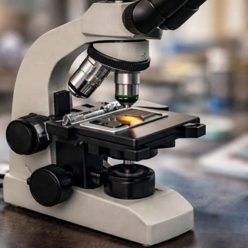
What equipment is typically used?
Basic mushroom spore microscopy usually involves:
A microscope
- Compound microscopes are commonly used
- Magnification ranges typically between 400x and 1000x
- Higher magnification reveals finer surface details
Microscope slides and coverslips
- Used to mount spores safely
- Help protect samples and lenses
Light source
- Transmitted light highlights spore shape and colour
- Adjustable lighting improves contrast
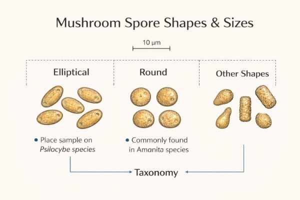
What do mushroom spores look like under a microscope?

Under magnification, mushroom spores may appear:
- Oval, elliptical, or spherical
- Smooth or textured
- Light to dark brown, purple-brown, or translucent
- Uniform or varied in size
These characteristics are often used to differentiate species and genera.
How spores are prepared for microscopy
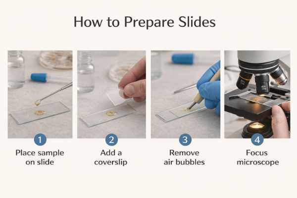
For educational and research purposes, spores are typically:
- Mounted on clean microscope slides
- Observed in small quantities
- Handled with care to avoid contamination
Different spore formats may be used depending on preference and availability.
For an overview of formats, see:
Spore Syringe vs Spore Print vs Spore Swab
Common observations recorded in spore microscopy
When examining spores, researchers often note:
- Size and shape
- Colour and opacity
- Surface texture
- Spore wall thickness
These observations contribute to fungal taxonomy and documentation.
Responsible handling during microscopy
Microscopy should always be conducted responsibly:
- Use spores only for lawful purposes
- Avoid unnecessary exposure or handling
- Store samples correctly after observation
- Follow all applicable laws and ethical guidelines
Understanding legality is an essential part of responsible study.
Legal context in the UK
While mushroom spores are legal to possess in the UK, cultivation of certain species is illegal.
Microscopy:
- Is a lawful, educational use
- Does not involve germination or cultivation
- Is widely recognised as legitimate scientific study
For full details, read:
Are Mushroom Spores Legal in the UK?
Frequently asked questions
Do I need an expensive microscope?
No. Many entry-level compound microscopes are suitable for observing spores at basic magnification levels.
What can mushroom spores tell you under a microscope?
Under a microscope, mushroom spores can reveal information about shape, size, colour, wall thickness, and surface texture, which are key characteristics used in fungal identification and taxonomy.
At what magnification are mushroom spores best observed?
Most mushroom spores can be clearly observed at 400× magnification, while finer surface details are typically examined at 1000× using oil immersion.
Do all mushroom spores look the same under a microscope?
No. Mushroom spores vary widely in shape, size, colour, and ornamentation depending on species and genus. These differences are important in distinguishing closely related fungi.
Can you identify a mushroom species using spores alone?
Spore characteristics can support identification, but they are rarely sufficient on their own. Accurate identification usually requires macroscopic features and ecological context in addition to microscopy.
What is spore ornamentation?
Spore ornamentation refers to the surface features of a spore, such as smooth walls, ridges, warts, or reticulated patterns, which may be visible at higher magnifications.
Why is oil immersion used at 1000× magnification?
Oil immersion improves optical resolution by reducing light refraction, allowing finer spore details to be seen more clearly at high magnification.
How large are typical mushroom spores?
Most mushroom spores range between 5–15 micrometres (µm) in length, though size varies widely depending on species.
Can mushroom spores be damaged during slide preparation?
Yes. Excessive pressure, contamination, or improper mounting mediums can distort or damage spores, making observation less accurate.
Can spores be damaged during microscopy?
Yes. Improper handling or contamination can affect samples, which is why careful preparation is important.
Is mushroom spore microscopy suitable for beginners?
Yes. Basic spore microscopy is a common entry point into mycology, provided that proper handling techniques and safety practices are followed.
Do spore colours under the microscope match spore print colours?
Not necessarily. Spore print colour reflects mass spore deposition, while individual spores under magnification may appear transparent, pale, or lightly pigmented.
How long do prepared spore slides last?
Temporary wet mounts are usually short-lived, while properly prepared permanent slides can last months or longer if sealed and stored correctly.
Is microscopy a valid reason to own spores?
Yes. Microscopy and educational study are common, legitimate uses for mushroom spores.
Final thoughts
Mushroom spore microscopy offers a fascinating look into fungal biology and supports responsible, educational engagement with mycology. With basic equipment and careful handling, spores can be studied safely and ethically.
Related learning resources
Written by Mycotown Editorial Team
The Mycotown Editorial Team is responsible for producing and reviewing educational and reference content across the site. Our focus is on accurate, responsible information relating to mushroom spores, microscopy, and lawful research practices. View author profile